Eyeball Printable
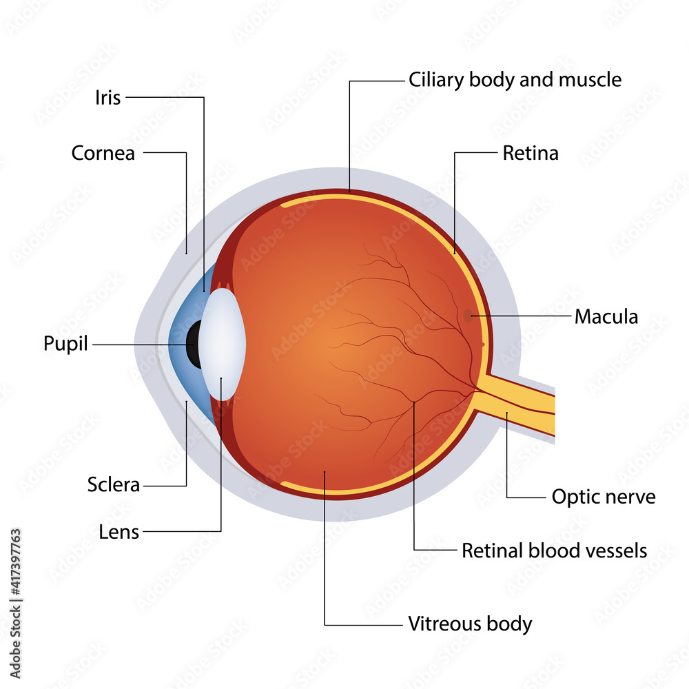
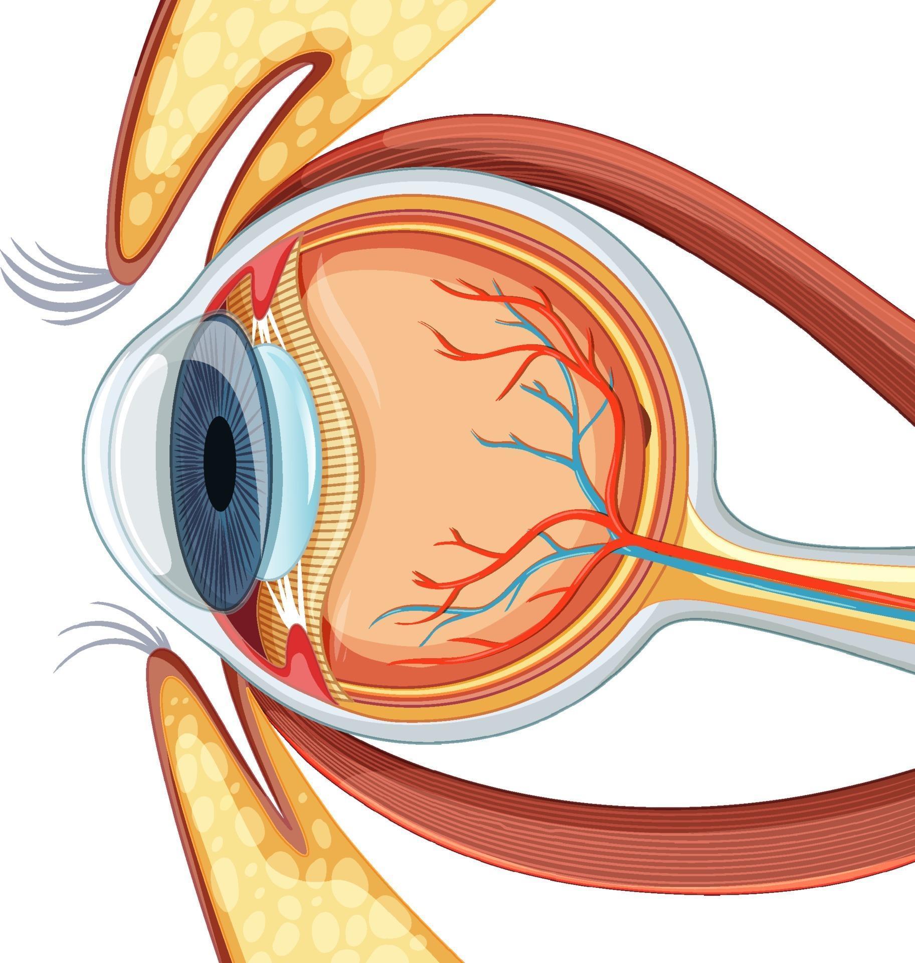
Eyeball Printable - The sclera is the thickest layer and is made of fibrous connective tissue that is. The sclera, choroid, and retina. The eyeball is a bilateral and spherical organ, which houses the structures responsible for vision. It consists of several layers and structures, each playing a. The outer sclera, middle choroid layer, and inner retina (fig. Learn everything about its anatomy and function at kenhub! It's composed of three primary layers: Know which parts of the eyeball are involved in accommodation. The eyeball is a round gelatinous organ that contains the actual optical apparatus. The eyeball houses the retina—an extremely. The eyeball is a bilateral and spherical organ, which houses the structures responsible for vision. The outer sclera, middle choroid layer, and inner retina (fig. The eye is cushioned within the orbit by pads of fat. In addition to the eyeball itself, the orbit contains the muscles that move the eye, blood vessels, and nerves. Learn everything about its anatomy and function at kenhub! Being able to describe different parts of a human eyeball. The sclera, choroid, and retina. It consists of several layers and structures, each playing a. Eyeball, spheroidal structure containing sense receptors for vision, found in all vertebrates and constructed much like a simple camera. The eyeball is a round gelatinous organ that contains the actual optical apparatus. The eyeball is a bilateral and spherical organ, which houses the structures responsible for vision. Know what a blind spot on the retina is, and where it’s located. Know which parts of the eyeball are involved in accommodation. The sclera, choroid, and retina. The outer sclera, middle choroid layer, and inner retina (fig. It's composed of three primary layers: In addition to the eyeball itself, the orbit contains the muscles that move the eye, blood vessels, and nerves. Eyeball, spheroidal structure containing sense receptors for vision, found in all vertebrates and constructed much like a simple camera. It is approximately 25 mm in diameter and sits snugly in the orbit where six muscles. Eyeball, spheroidal structure containing sense receptors for vision, found in all vertebrates and constructed much like a simple camera. In its wall, the eyeball has three layers: The eye is cushioned within the orbit by pads of fat. The bulb of the eye (bulbus oculi; It's composed of three primary layers: Learn everything about its anatomy and function at kenhub! In addition to the eyeball itself, the orbit contains the muscles that move the eye, blood vessels, and nerves. The eyeball houses the retina—an extremely. Know which parts of the eyeball are involved in accommodation. The bulb of the eye (bulbus oculi; The eyeball is a bilateral and spherical organ, which houses the structures responsible for vision. The eyeball is a round sensory organ that enables us to see. The sclera, choroid, and retina. The eyeball is a round gelatinous organ that contains the actual optical apparatus. It consists of several layers and structures, each playing a. In its wall, the eyeball has three layers: The eyeball is a complex and highly specialized organ that is responsible for capturing and processing visual information. It consists of several layers and structures, each playing a. The outer sclera, middle choroid layer, and inner retina (fig. Learn everything about its anatomy and function at kenhub! It is approximately 25 mm in diameter and sits snugly in the orbit where six muscles control its. The eye is cushioned within the orbit by pads of fat. The outer sclera, middle choroid layer, and inner retina (fig. The eyeball is a round gelatinous organ that contains the actual optical apparatus. The eyeball is a complex and highly specialized. The outer sclera, middle choroid layer, and inner retina (fig. Learn everything about its anatomy and function at kenhub! The eyeball is a round gelatinous organ that contains the actual optical apparatus. In addition to the eyeball itself, the orbit contains the muscles that move the eye, blood vessels, and nerves. Being able to describe different parts of a human. The sclera is the thickest layer and is made of fibrous connective tissue that is. The human eyeball is an intricate structure, roughly spherical in shape, with a diameter of about 24 millimeters. It is approximately 25 mm in diameter and sits snugly in the orbit where six muscles control its. The bulb of the eye (bulbus oculi; In addition. The eye is cushioned within the orbit by pads of fat. The eyeball is a round gelatinous organ that contains the actual optical apparatus. The eyeball is a bilateral and spherical organ, which houses the structures responsible for vision. It consists of several layers and structures, each playing a. The bulb of the eye (bulbus oculi; The eyeball is a bilateral and spherical organ, which houses the structures responsible for vision. The outer sclera, middle choroid layer, and inner retina (fig. Know which parts of the eyeball are involved in accommodation. In addition to the eyeball itself, the orbit contains the muscles that move the eye, blood vessels, and nerves. The eyeball is a round sensory organ that enables us to see. It's composed of three primary layers: It consists of several layers and structures, each playing a. The eyeball is a complex and highly specialized organ that is responsible for capturing and processing visual information. The eye is cushioned within the orbit by pads of fat. Eyeball), or organ of sight, is contained in the cavity of the orbit, where it is protected from injury and moved by the ocular muscles. The eyeball houses the retina—an extremely. Know what a blind spot on the retina is, and where it’s located. It is approximately 25 mm in diameter and sits snugly in the orbit where six muscles control its. The human eyeball is an intricate structure, roughly spherical in shape, with a diameter of about 24 millimeters. The eyeball is a round gelatinous organ that contains the actual optical apparatus. The sclera, choroid, and retina./GettyImages-1128675065-e4bac15b0f39449dba31f25f1020bc8a.jpg)
Eyeball
Eyeball Diagram Labeled Eyeball Anatomy
Eyeball Structure, Function & Muscles Britannica
![]()
Eyeball Anatomy

Eyeball Parts Anatomy

Structure Anatomy Human Eye Realistic Eyeball Vector vrogue.co
Eyeball
Premium Photo Human eye isolated on a white background realistic
Eyeball
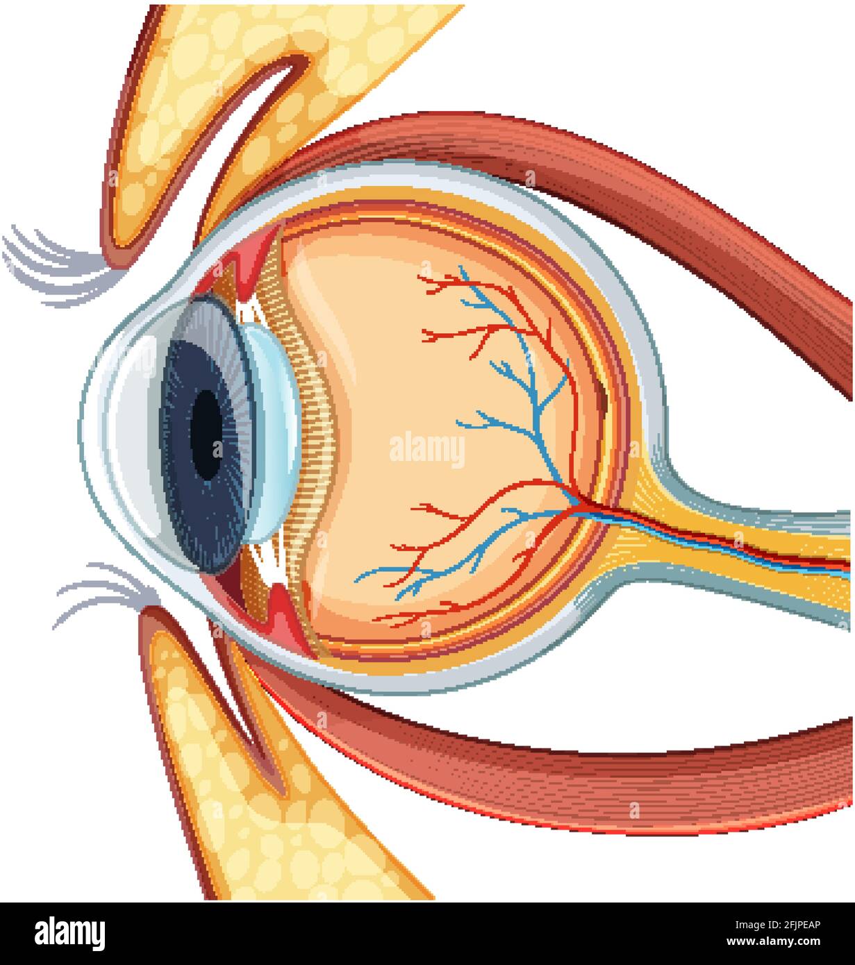
Diagram of human eyeball anatomy illustration Stock Vector Image & Art
Learn Everything About Its Anatomy And Function At Kenhub!
Being Able To Describe Different Parts Of A Human Eyeball.
In Its Wall, The Eyeball Has Three Layers:
The Sclera Is The Thickest Layer And Is Made Of Fibrous Connective Tissue That Is.
Related Post: